Diagram Cross Section Of A Bone : Medical Diagram Of The Structure Of The Inside Cross Section Of The Tooth Stock Vector Illustration Of Medical Education 125924960 - I am not an expert on this subject, so i was wondering if anyone could put their input on it seems confusing and misleading.
Diagram Cross Section Of A Bone : Medical Diagram Of The Structure Of The Inside Cross Section Of The Tooth Stock Vector Illustration Of Medical Education 125924960 - I am not an expert on this subject, so i was wondering if anyone could put their input on it seems confusing and misleading.. There are trabeculae in spongy bone which gives its sponge like appearance. Bone cross section diagram ipad folio cases. Скелет человека/ anatomy of the bone system. Two types of bone tissues in cross section of a long bone : The centroidal distance, c, is the distance from the centroid of a cross section to the extreme fiber.
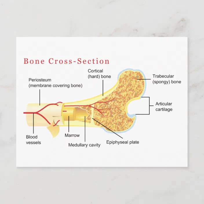
For example, to read this diagram literally, since the cartilage can be seen inside the cutaway section of bone, it. Cross section diagrams a cross section diagram is if you would take a knife and cut through one side of a diagram to see the inside and outside in one picture. They build the entire picture, improve your understanding, consolidate the information and facilitate recall. Explaned distal and proximal epiphysis. Diagram with articular cartilage, marrow, spongy bone, medullary cavity, endosteum, diaphysis, and periosteum. can be used for personal and commercial purposes.
Mammal Bones 101 The Magic Of Bone Growth Diarthrodial Joints from www.earthlife.net This article covers the anatomy of the spinal cord including its structure tracts and function. Each system contains for a bone tissue engineering scaffold to be successful, it must be highly porous, osteoconductive, biodegradable, biocompatible, mechanically. Blood vessels and nerves enter the bone through the. U0026quot bone cross section u0026quot for radius digital science on behance. Bone contains a relatively small number of cells entrenched in a matrix of collagen fibers that provide a surface for inorganic salt crystals to adhere. Diagram with articular cartilage, marrow, spongy bone, medullary cavity, endosteum, diaphysis, and periosteum. can be used for personal and commercial purposes. Cross section diagrams a cross section diagram is if you would take a knife and cut through one side of a diagram to see the inside and outside in one picture. Bone is found in the shafts of long bone and consists of various cylindrical units named as haversian system 47.
The large dark spots are passages for blood vessels and nerves.
Diagram with articular cartilage, marrow, spongy bone, medullary cavity, endosteum, diaphysis, and periosteum. Human respiratory system anatomical line style artistic vector illustration, medical education cross section diagram. The surface features of bones vary considerably, depending on the function and location in the body. Скелет человека/ anatomy of the bone system. The large dark spots are passages for blood vessels and nerves. In the last decade, considerable technological improvements have been made to repair damaged bones and tissue, such as bone cross sections with implants for microscopic examinations. Diagram with articular cartilage, marrow, spongy bone, medullary cavity, endosteum, diaphysis, and. Diagram of blood and nerve supply to bone. Whereas a long bone has only one layer of compact bone (see fig 1). Diagram of a cross section of the coiled cochlea. Spongy bone and compact bone. There are trabeculae in spongy bone which gives its sponge like appearance. Health, bones, one object, vein, human skeleton, artery, cavity, skeletal system, nerve, compact, human bone, human tissue, human nervous system, marrow, spongy bone, porous, connective tissue, spongy, human artery, cancellous bone, diaphysis.
Diagram with articular cartilage, marrow, spongy bone, medullary cavity, endosteum, diaphysis, and periosteum. can be used for personal and commercial purposes. Select from premium cross section of bone images of the highest quality. The large dark spots are passages for blood vessels and nerves. This article covers the anatomy of the spinal cord including its structure tracts and function. For example, to read this diagram literally, since the cartilage can be seen inside the cutaway section of bone, it.
Bone Cross Section Diagram Postcard Zazzle Com from rlv.zcache.com Health, bones, one object, vein, human skeleton, artery, cavity, skeletal system, nerve, compact, human bone, human tissue, human nervous system, marrow, spongy bone, porous, connective tissue, spongy, human artery, cancellous bone, diaphysis. These bone cells have long branching arms (d) which lets them communicate with. Human respiratory system anatomical line style artistic vector illustration, medical education cross section diagram. These bone cells (described later) cause the bone to grow, repair, and remodel throughout life. Explaned distal and proximal epiphysis. Bone cross section diagram ipad folio cases. They build the entire picture, improve your understanding, consolidate the information and facilitate recall. For example, to read this diagram literally, since the cartilage can be seen inside the cutaway section of bone, it.
Diagram with articular cartilage, marrow, spongy bone, medullary cavity, endosteum, diaphysis, and periosteum. can be used for personal and commercial purposes. Explaned distal and proximal epiphysis. For example, to read this diagram literally, since the cartilage can be seen inside the cutaway section of bone, it. Cross section diagrams a cross section diagram is if you would take a knife and cut through one side of a diagram to see the inside and outside in one picture. Скелет человека/ anatomy of the bone system. Two types of bone tissues in cross section of a long bone : Diagram with articular cartilage, marrow, spongy bone, medullary cavity, endosteum, diaphysis, and periosteum. Label the parts of a long bone. The centroidal locations of common cross sections are well documented, so it is typically not necessary to calculate the location with the equations above. Diagram of the long bone. U0026quot bone cross section u0026quot for radius digital science on behance. Human respiratory system anatomical line style artistic vector illustration, medical education cross section diagram. They build the entire picture, improve your understanding, consolidate the information and facilitate recall.
Cross section diagrams a cross section diagram is if you would take a knife and cut through one side of a diagram to see the inside and outside in one picture. These bone cells have long branching arms (d) which lets them communicate with. These bone cells (described later) cause the bone to grow, repair, and remodel throughout life. The surface features of bones vary considerably, depending on the function and location in the body. The large dark spots are passages for blood vessels and nerves.
Bone Histology Of The Caudata A Mid Diaphyseal Cross Section In Download Scientific Diagram from www.researchgate.net Diagram with articular cartilage, marrow, spongy bone, medullary cavity, endosteum, diaphysis, and periosteum. Compact bone is the outer layer and the spongy bone forms the inner layer. Diagram with articular cartilage, marrow, spongy bone, medullary cavity, endosteum, diaphysis, and periosteum. can be used for personal and commercial purposes. As shown in figure 2. We have weaker bones than our hunter. These bone cells have long branching arms (d) which lets them communicate with. File human leg bones labeled svg. How to draw the diagram of cross section of a leaf class x.
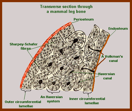
Diagram of cross section of bone.
Two types of bone tissues in cross section of a long bone : Explaned distal and proximal epiphysis. Health, bones, one object, vein, human skeleton, artery, cavity, skeletal system, nerve, compact, human bone, human tissue, human nervous system, marrow, spongy bone, porous, connective tissue, spongy, human artery, cancellous bone, diaphysis. We have weaker bones than our hunter. Spongy bone and compact bone. Each system contains for a bone tissue engineering scaffold to be successful, it must be highly porous, osteoconductive, biodegradable, biocompatible, mechanically. Cross section diagrams a cross section diagram is if you would take a knife and cut through one side of a diagram to see the inside and outside in one picture. Jump to navigation jump to search. Function of bone bone is a living, metabolically active and highly organized tissue consisting of a. So what im going to do is jump right to the. Blood vessels and nerves enter the bone through the. We can see there are two layers of compact bone here. As the names suggest compact bone looks compact and the spongy bone looks like skull bone is a flat bone.
Bone contains a relatively small number of cells entrenched in a matrix of collagen fibers that provide a surface for inorganic salt crystals to adhere cross section of a bone. For example, to read this diagram literally, since the cartilage can be seen inside the cutaway section of bone, it.


0 Komentar